ClinicalKey
| Site: | IMU University Library Portal |
| Course: | Subject Based e-Resources |
| Book: | ClinicalKey |
| Printed by: | Guest user |
| Date: | Thursday, 23 July 2026, 4:06 AM |
Description
1. eBooks
|
Gray's Anatomy, Forty-Third Edition Standring, Susan, MBE, PhD, DSc, FKC, Hon FAS, Hon FRCS © 2026, Elsevier Limited. All rights reserved. |
Atlas of Human Anatomy, Eighth Edition Netter, Frank H., MD Copyright © 2022 by Elsevier Inc. |
|
Fitzgerald's Clinical Neuroanatomy and Neuroscience, Eighth Edition Mtui, Estomih, MD Copyright © 2021 by Elsevier Limited. All rights reserved. Previous editions copyrighted 2004, 1997. |
More Info about ClinicalKey eBooks, click HERE
2. Videos
Anatomy Videos - ClinicalKey |
|
|
Video 1 - Anatomical Arthroscopic Anterior Talofibular Ligament and Calcaneofibular Ligament Reconstruction Using an Autogenic Hamstring Tendon: Safe Creation of Anatomical Fibular Tunnel Arthroscopy Techniques. Higashiyama, Reiji, M.D., Ph.D.; Aikawa, Jun, M.D., Ph.D.; Iwase, Dai, M.D.; Takamori, Yasuyuki, M.D., Ph.D.; Watanabe, Eiichiro, M.D., Ph.D.; Takaso, Masashi, M.D., Ph.D.. Published March 1, 2019. Surgical technique for anatomical arthroscopic ATFL and CFL reconstruction using an autogenic hamstring tendon for a safe creation of an anatomical fibular tunnel. This surgery is performed under general anesthesia with the patient in the supine p...
|
Video 1 - Anatomical Arthroscopic Anterior Talofibular Ligament Repair and Reconstruction Using a Free Tendon Arthroscopy Techniques. Higashiyama, Reiji, M.D., Ph.D.; Sekiguchi, Hiroyuki, M.D., Ph.D.; Takata, Ken, M.D.; Katagiri, Akira, M.D.; Inoue, Gen, M.D., Ph.D.; Takaso, Masashi, M.D., Ph.D.. Published January 1, 2020. Surgical technique for anatomic arthroscopic anterior talofibular ligament (ATFL) repair and reconstruction using a free tendon in the right ankle. This surgical procedure is performed with the patient under general anesthesia in the supine positi...  |
|
Video 1 - Anatomic knotless distal triceps tendon repair. Arthroscopy Techniques. Kevin O’Donnell, MD; David L. Rubenstein, MD; Michael G. Ciccotti, MD Ph.D.. Published 2018. The patient is placed in the prone or lateral decubitus position with the elbow flexed to 90 degrees. A midline curvilinear incision is made over the posterior aspect of the elbow. The incision is curved to the radial side of the tip of the olecranon, which prevents the healed incision from being in the direct area of contact ...
|
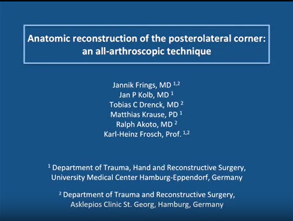
Video 1 - Anatomic Reconstruction of the Posterolateral Corner: An All-Arthroscopic Technique Arthroscopy Techniques. Frings, Jannik, M.D.; Kolb, Jan P., M.D.; Drenck, Tobias C., M.D.; Krause, Matthias, M.D.; Alm, Lena, M.D.; Akoto, Ralph, M.D.; Frosch, Karl-Heinz, M.D.. Published February 1, 2019. The posterolateral corner consists of the lateral collateral ligament (LCL) and the popliteus complex, which comprises the popliteus muscle tendon unit and the arcuate complex. The popliteus complex, especially the arcuate complex, stabilizes the ...
|
| More Info about ClinicalKey Videos, click HERE | |


3. Images
|
|
|
|
|
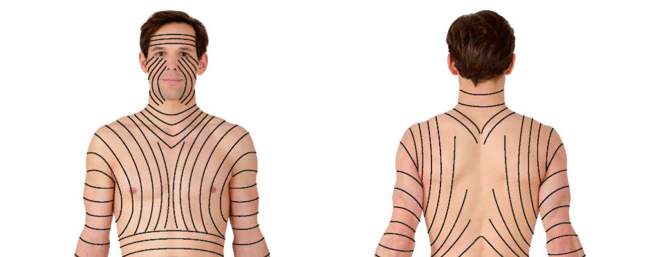 General AnatomyFig. 1.11a and b:Segmental innervation of the skin (dermatomes). a Ventral view, b dorsal view. [L126] A dermatome is an area of skin innervated autonomously by the sensory fibres of a spinal cord nerve (spinal nerves, → Fig. 1.45). Each spinal nerve can thus be a... Sobotta Atlas of Anatomy, Vol.1, 16th ed., English/Latin. Paulsen, Friedrich.. Published January 1, 2018. Pages 1-54. © 2018. |
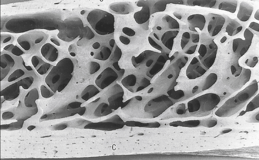 Anatomy of the musculoskeletal systemFig. 5.8:A vertical section 2 cm below the anterosuperior border of the iliac crest (female, 42 years). The cancellous bone consists of intersecting curved plates and struts. Osteonal (Haversian) canals can just be seen in the two cortices...
Gray's Anatomy. Mendelson, Bryan C; Wong, Chin-Ho.. Published January 1, 2021. Pages 85-126. © 2021. |
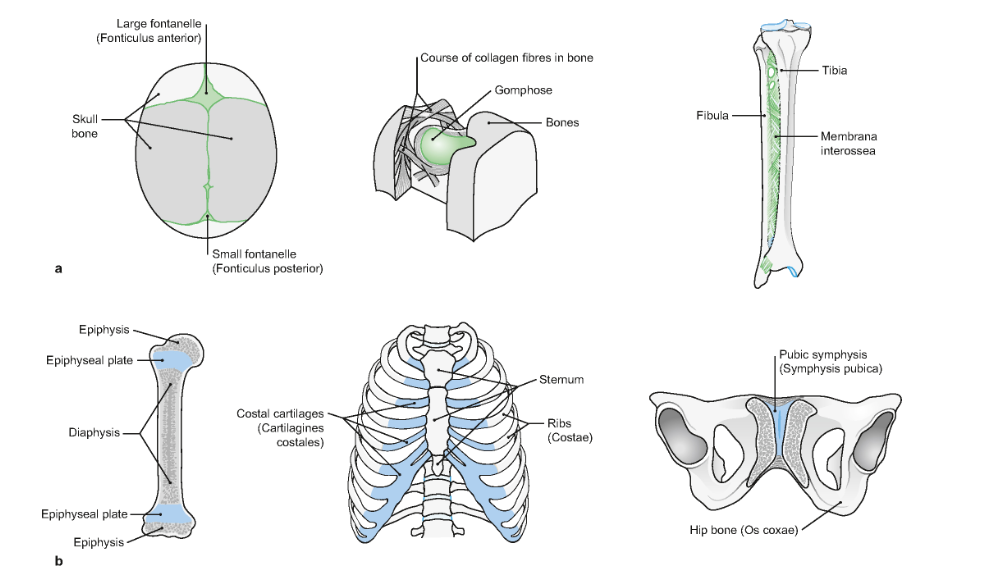
 General AnatomyFig. 1.19a:Structure of a long tubular bone, os longum; section through the proximal part of the right thigh bone (femur) of an adult. In the area of the diaphysis (bone shaft) the periosteum (bone membrane) is raised and to the side; dorsal view. a Macrosco... Sobotta Atlas of Anatomy, Vol.1, 16th ed., English/Latin. Paulsen, Friedrich.. Published January 1, 2018. Pages 1-54. © 2018. |
More Info about ClinicalKey Images, Click HERE