
ClinicalKey
3. Images
|
|
|
|
|
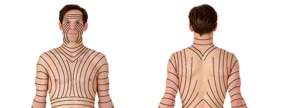 General AnatomyFig. 1.11a and b:Segmental innervation of the skin (dermatomes). a Ventral view, b dorsal view. [L126] A dermatome is an area of skin innervated autonomously by the sensory fibres of a spinal cord nerve (spinal nerves, → Fig. 1.45). Each spinal nerve can thus be a... Sobotta Atlas of Anatomy, Vol.1, 16th ed., English/Latin. Paulsen, Friedrich.. Published January 1, 2018. Pages 1-54. © 2018. |
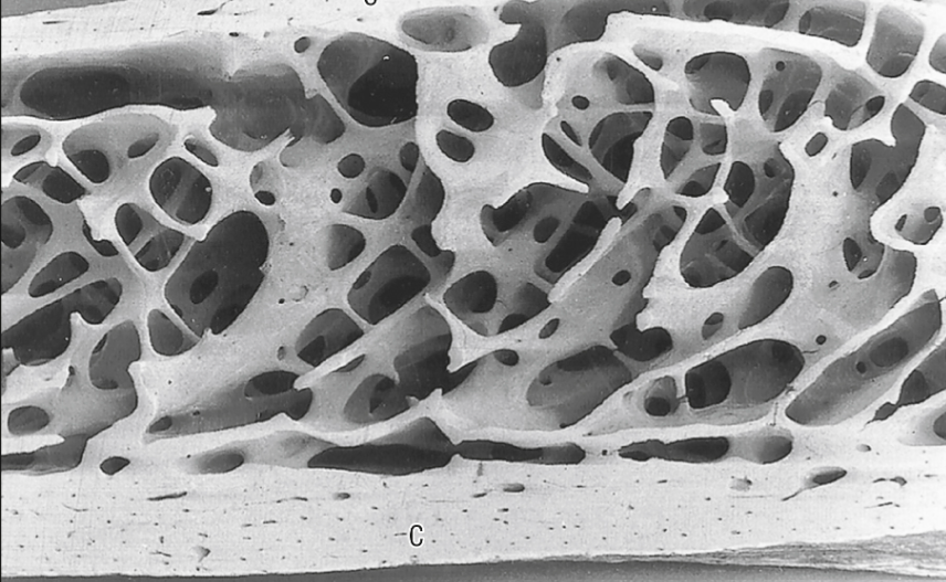 Anatomy of the musculoskeletal systemFig. 5.8:A vertical section 2 cm below the anterosuperior border of the iliac crest (female, 42 years). The cancellous bone consists of intersecting curved plates and struts. Osteonal (Haversian) canals can just be seen in the two cortices...
Gray's Anatomy. Mendelson, Bryan C; Wong, Chin-Ho.. Published January 1, 2021. Pages 85-126. © 2021. |
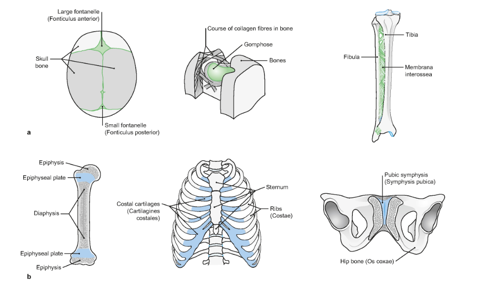
 General AnatomyFig. 1.19a:Structure of a long tubular bone, os longum; section through the proximal part of the right thigh bone (femur) of an adult. In the area of the diaphysis (bone shaft) the periosteum (bone membrane) is raised and to the side; dorsal view. a Macrosco... Sobotta Atlas of Anatomy, Vol.1, 16th ed., English/Latin. Paulsen, Friedrich.. Published January 1, 2018. Pages 1-54. © 2018. |
More Info about ClinicalKey Images, Click HERE