
Thieme
2. Images/Videos/Audio
|
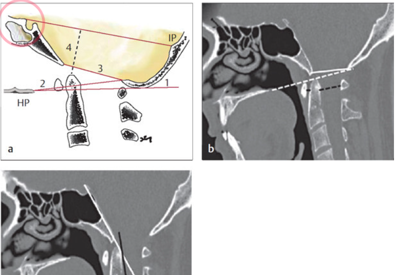
Differential Diagnosis in Neurology and Neurosurgery: A Clinician’s Pocket Guide Source: Tsementzis S, ed. Differential Diagnosis in Neurology and Neurosurgery: A Clinician’s Pocket Guide. 2nd Edition. 2019. Fig. 2.23 (a) Radiographic criteria for basilar impression. 1: McGregor’s line between hard palate (HP) and the lowest point of occiput. Basilar impression is present if the dens protrudes more than 5 mm above this line. 2: Chamberlain’s line between hard palate and opisthion. Positive diagnosis if dens protrudes more than 2.5 mm above line. |
Thieme Test Prep for the USMLE®: Clinical Anatomy Q&A Source: Riede U, Werner M. Color Atlas of Pathology. 1st Edition. Thieme; 2004. Source: Hankin M, Morse D, Venuti J et al., ed. Thieme Test Prep for the USMLE®: Clinical Anatomy Q&A. 1st Edition. 2019 |
|
Differential Diagnosis in Neurology and Neurosurgery: A Clinician’s Pocket Guide. Fig. 2.23 (a) Radiographic criteria for basilar impression. 1: McGregor’s line between hard palate (HP) and the lowest point of occiput. Basilar impression is present if the dens protrudes more than 5 mm above this line. 2: Chamberlain’s line between hard palate and opisthion. Positive diagnosis if dens protrudes more than 2.5 mm above line. Source: Tsementzis S, ed. Differential Diagnosis in Neurology and Neurosurgery: A Clinician’s Pocket Guide. 2nd Edition. 2019. |

More Details of Thieme Images/Videos/Audio - Pathology, click HERE